Andrea (name changed) is in her 40’s and runs a busy household. For a few months, she had been waking up with a headache. Not daily at first, though recently they had become more frequent. The headache would resolve after being up a while, but recently it has lingered through the day.
She scheduled a visit with her family doctor. There, she described a frontal, throbbing, and, at times, sharp headache. The past couple of weeks, she reported, episodes of numbness and weakness in the right hand and dizziness when changing positions.
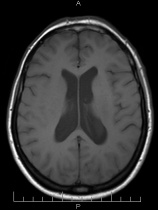
A neurological examination revealed moderate difficulty walking with one foot in front of the other. Otherwise, the exam was normal. Her provider ordered an MRI of the brain which revealed the ventricles (spinal fluid spaces inside the brain) to be somewhat larger than normal – a condition called hydrocephalus (Figure 1). Something was blocking the flow of spinal fluid through the ventricles, but what could it be?

Figure 1 – Cross-section MRI showing enlargement of the ventricles (dark fluid filled spaces) in the center of the brain.
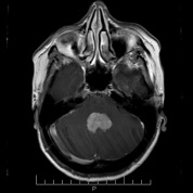
In the side view (sagittal) MRI, a tumor could be seen inside of the fourth ventricle, the fluid space between the brainstem and the cerebellum (Figure 2). A cross section MRI scan (Figure 3), performed after she had received received intravenous contrast (“dye” material given through a vein), clearly outlined the tumor (Figure 3). It was blocking blocking cerebrospinal fluid (CSF) flow and enlarging the ventricle. This explained why the ventricles in the upper brain looked too big.

Figure 3 – Cross section view of the lower brain area. The tumor is easily after intravenous contrast was given.
Comment
There are various treatments for brain tumors. Some tumors are first diagnosed with a needle biopsy, while others may be treated with specialized radiation called radiosurgery. However, in many cases, surgery is required to remove the tumor. In Andrea’s case, the tumor was blocking CSF flow. If the blockage became worse, which was very likely since the tumor would keep growing, it could render her unconscious and jeopardize her life. A needle biopsy would not resolve the CSF blockage.
Surgery
After careful discussions with her and her husband, she was readied for surgery. The plan was to reach the tumor through a cranial opening (craniotomy) at the back of the head. Once the membrane surrounding the brain was exposed and opened, a path would be developed between the two halves of the lower cerebellum (called the cerebellar tonsils). With gentle separation of the cerebellar tonsils, the lower part of the tumor came into view (Figure 4).

Figure 4 – Microphotograph during surgery showing the lower portion of the tumor between the cerebellar tonsils.
After a biopsy was obtained, the tumor was removed through microsurgery using the neurosurgical microscope, microsurgical instruments, and an ultrasonic aspirator – an instrument whose tip vibrates at ultrasonic frequencies and morselize (break-down) the cells. (This tool has replaced the laser that came into use in the 1980’s, although there are still a few applications for it today). Using the gentle action of the ultrasonic aspirator and microsurgical dissection, the tumor was steadily removed. Inspection of the floor of the fourth ventricle showed no residual tumor (Figure 5).
Andrea stayed in the hospital for … days and was then discharged home with her family. She steadily resumed her normal activities. Follow-up brain MRI scans have shown no residual tumor (Figure 6). Happily, she has recovered completely and is back to her busy life.
Comment
Tumors growing within the cerebral ventricles are rare in the adult population. In one study of 73 persons with intraventricular tumors, the types of tumor found included:
- medulloblastoma
- ependymoma
- epidermoid
- cystercircosis
- astrocytoma
- subependymoma
Other tumors found within the ventricles are a choroid plexus papillomas and meningiomas. A metastasis from a tumor elsewhere in the body can also develop within the ventricles.
The pathological examination of Andrea’s tumor revealed it to be a choroid plexus papilloma. Follow-up MRI scans within the first year following surgery have shown no tumor. The chances of the tumor coming back from unseen cells buried within the tissues are about 10%. MRI scans will be performed on a regular basis for a few years.
REFERENCE
Intraventricular mass lesions.
Morrison G, Sobel DF, Kelley WM, Norman D.
Radiology. 1984 Nov;153(2):435-42.


